
Thoracoabdominal Aortic Surgery
Background
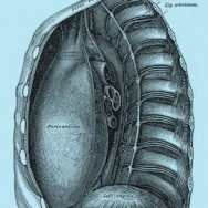
The aorta is the large artery that leaves the heart and provides oxygen-rich blood throughout the body. It first travels upwards (the ascending aorta), before bending backwards (the arch) and downwards to supply the lower parts of the body (the descending aorta). The descending aorta has a segment above the diaphragm (the thoracic part) and below the diaphragm (the abdominal part).
Many conditions can cause the descending aorta to dilate (widen), dissect (tear), or rupture. These are often life-threatening and require emergency surgery to repair them. The conditions that can lead to this include atherosclerosis (fat and calcium buildup in the arteries), hypertension (high blood pressure), genetic conditions (such as Marfan Syndrome an bicuspid valve aortopathy), connective tissue disorders (such as Ehler-Danlos disorder, scleroderma) and direct trauma.
About the surgery
First the aorta is accessed via a large incision extending from the left side of the chest and over the abdomen. Then the patient is put on the heart-lung machine. The circulation through the descending aorta must then be stopped so that the diseased segment can be removed, and a synthetic replacement graft inserted. Blood flow to the heart, brain and rest of the body is maintained using the heart-lung machine.
The descending aorta supplies blood to the spinal cord, though since the circulation is stopped during the repair a number of precautions are taken to prevent spinal cord injury. Firstly, some of the artery branches to the spine are reconnected to the graft so that blood can flow through them after again the operation. Secondly, a separate heart-lung machine circuit is used to supply blood to the bottom part of the descending aorta. Other things that are used include draining some of the spinal cord fluid to lower the pressure in the spinal cord and increase the blood supply, lowering the patient’s temperature, and comprehensive monitoring of spinal cord function (sensory and motor evoked potentials). Sydney Heart & Lung Surgeons are experienced at this complex operation, and take every precaution to prevent neurological or spinal cord injury.
Recovery period
Patients usually stay in the intensive care unit for 1 – 2 days for monitoring, and then in the nursing unit for 6-8 days. Two to three tubes stay in the chest to drain fluid from around the aorta, which are usually removed 1 – 3 days after the surgery. Full recovery usually takes about 3 months. Your surgeon and nurse case manager will provide specific guidelines for your recovery and return to work.
Risks of the surgery
Because of the complexity of these operations there are some risks involved. These include stroke, bleeding, lung failure, kidney failure, damage to the bowel, and death. Your surgeon will discuss these risks with you, as they are balanced with the risks of not intervening.
The rate of these complications is lower in experienced centres. Surgeons at Sydney Heart & Lung Surgeons have performed many of these procedures with excellent results, and have published these results in international peer-reviewed journals.
For more information please visit:
Cleveland Clinic Thoracic Aortic Aneurysm
All patients should consult their cardiothoracic surgeon for specific information about their medical condition and surgery.